Dental X-rays

What are the benefits of a dental x-ray exam?
Radiographic or X-ray examinations provide your dentist with an important tool to see the structures of your mouth that can not be seen visually, such as the roots of the teeth and the bone that surrounds them.
X-rays can locate cavities and other problems like abscesses, cysts, tumours, and impacted or missing teeth. Periodontal (gum) disease and its severity can be determined.
How do dental x-rays work?
When X-rays pass through your mouth during a dental examination, more X-rays are absorbed by the denser parts (such as teeth and bone) than by soft tissues (such as cheeks and gums) before striking the film. This creates an image on the radiograph. Teeth appear lighter because fewer X-rays penetrate to reach the film. Cavities and gum disease appear darker because of more X-ray penetration. The interpretation of these X-rays allows the dentist to safely and accurately detect hidden abnormalities.
With digital dental X-rays, we can quickly take images of your teeth and mouth, enabling us to detect tooth decay and cavities earlier, allowing us to set up preventive treatment plans that can help small issues from becoming big problems. The latest technology allows dentists to enhance and magnify your images, with the ability to zoom in/focus on specific teeth or areas of the mouth – showing a much closer and clear shot.

Compared to traditional dental X-ray’s, exposure to X-ray radiation is reduced by as much as 80 per cent through digital dental X-rays. And, because the pictures are downloaded to a computer immediately, our dental professionals are provided with immediate access to your X-rays.
How often should x-rays be taken?
How often dental X-rays (radiographs) should be taken depends on the patient's individual health needs. Your dentist will review your history, examine your mouth and then decide whether you need radiographs and what type.
.
The schedule for needing radiographs at recall visits varies according to your age, risk for disease and signs and symptoms. Recent films may be needed to detect new cavities, or to determine the status of gum disease or for evaluation of growth and development. Children may need X-rays more often than adults. This is because their teeth and jaws are still developing and because their teeth are more likely to be affected by tooth decay than those of adults.
Should I be concerned about exposure to radiation?

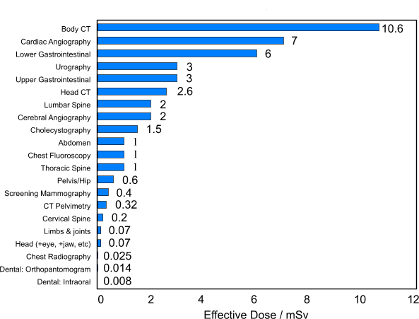
Typical values of dose for various medical and dental x-ray procedures
In the above chart, you can see the relative amounts of exposure typical diagnostic radiographs produce. The dental intra-oral films are periapical or bitewing type X-rays. While you can see above that films produce a relatively small amount of exposure, dentists are sensitive to patients’ concerns about exposure to radiation.
Your dentist has been trained to prescribe radiographs when they are appropriate and to tailor radiographic schedules to each patient's individual needs and therefore minimise the patients exposure.
Why do the dentist and assistant leave the room when they take x-rays of my teeth?
If your dentist or dental staff member does not leave the room, he or she will be exposed to radiation many times a day. Although the amount of radiation he or she receives each time is quite small, over time it could add up to a large amount.
Types of dental x-rays taken
Bitewing X-rays show the upper and lower back teeth and how the teeth touch each other in a single view. These X-rays are used to check for decay between the teeth and to show how well the upper and lower teeth line up. They also show bone loss when severe gum disease or a dental infection is present.
Periapical X-rays show the entire tooth, from the exposed crown to the end of the root and the bones that support the tooth. These X-rays are used to find dental problems below the gum line or in the jaw, such as impacted teeth, abscesses, cysts, tumours, and bone changes linked to some diseases.
Panoramic (OPG) X-rays show a broad view of the jaws, teeth, sinuses, nasal area, and jaw joints. These X-rays do not find cavities. These X-rays do show problems such as impacted teeth, bone abnormalities, cysts, solid growths (tumours), infections, and fractures.


If you have any further concerns about dental x-rays, please call or contact us through our website.
Why are dental x-rays important?
An example of why your dentist cannot rely on just a visual examination of your teeth is highlighted in the case shown below. The use of dental x-rays can help your dentist diagnose and treat conditions and problems invisible to the naked eye.
This preoperative photo of a molar (A), reveals no clinically apparent decay other than a small spot on the enamel surface. In fact, decay could not be detected with a dental probe.
Radiographic evaluation, (B), however, revealed an extensive region of demineralisation within the dentine (arrows) of the front half of the tooth.
When a bur was used to remove the enamel overlying the decay, (C), a large hollow was found within the tooth.

After all of the decay had been removed, (D), the pulp chamber had been exposed and most of the front half of the tooth was either missing or poorly supported. Root canal treatment was necessary to save the tooth.
How to find us
Modern Dentistry
Level 1 City Walk Centre
City Walk
Civic
Canberra
ACT 2601
Above King O'Malleys Pub
Work: 02 6247 8400
Opening Hours
Modern Dentistry
Monday - 8:30 am - 5:30 pm
Tuesday - 8:30 am - 5:00 pm
Wednesday - 8:30 pm - 5:30 pm
Thursday - 8:30 am - 5:30 pm
Friday - 8:00 am - 3:30 pm
Saturday - Closed
Sunday - Closed
